The Challenge
UCSF wanted to study the impact of orthodontics treatment on the temporomandibular joints (TMJ), but it couldn’t find an efficient, easy tool to view, manipulate and measure the joints in 3D based on DICOM data.
The Solution
Stratovan’s Checkpoint™ for medical imaging - a comprehensive set of state-of-the-art 3D shape analysis and visualization tools, provides better landmark-based measurement capabilities for a deeper understanding of 3D anatomic structures.
Goal: Understanding Joint Movement
Millions of people wear braces to correct orthodontics issues, or have maxillofacial surgery to improve poor occlusion. But until recently, little was known about the impact of such treatments on the temporomandibular joints (TMJ). “Braces change how the lower jaw occludes with the upper, and moves teeth, which moves the jaw in the TMJ,” says Dr. Renie Ikeda, DDS, who practices in San Francisco, California. “Sometimes this affects the TMJ and can cause pain in the jaw for a patient.”
Little research has been done to measure how the TMJ changes due to treatment, or to quantify the pain some patients experience after treatment. Dr. Ikeda, working with Dr. Arthur J. Miller, Professor of the Orofacial Sciences and Physiology Depart-ment at the University of California, San Francisco, decided to explore the issue.

“I wanted to do joint congruence analysis to see changes in spatial relations between the fossa and condyle after TMJ surgery, and objectively quantify those changes,” says Dr. Ikeda.
“However, I wanted to do more than just visually compare images taken of a patient over time to measure changes in the joint and shapes.”
Existing tools? Enough to cause jaw pain
To look at landmark features, several conventional methods exist that use multi-plane views. “I was planning to use a 2D structural analysis tool to look through the joint and do a linear measurement,” says Dr. Ikeda. “But, this is time-consuming and not very accurate.”
Another obstacle for Dr. Ikeda was the fact that conventional spatial analysis systems require the use of multiple software to download background and image files. “I wasn’t satisfied with the spatial analysis and imaging tools on the market, says Dr. Ikeda. ”I needed one comprehensive 3D measurement and analysis tool that would allow me to precisely measure the gap between the two joints that make up the TMJ, and see how it changes during treatment, by easily manipulating and comparing 3D images over time.”
Checking out Checkpoint

Then Dr. Ikeda learned about and started using Checkpoint™ from Stratovan Corporation. A set of state-of-the-art 3D shape analysis and visualization tools, Checkpoint gives healthcare professionals better landmark-based tools that facilitate a deeper understanding of 3D anatomic structures.
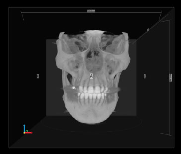
It provides 3D views of CT, MRI, PET and other 3D scans from a variety of modalities - including 3D surface scans - and enables the efficient collection of thousands of landmark points to provide precise analyses of complex 3D shapes.
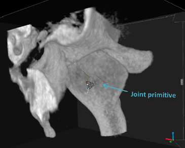
Checkpoint provides full 3D reconstruction to easily slice through for orthogonal and oblique views. It can load both surface meshes and volumetric DICOM scans, and extract multiple surfaces from a DICOM volume. A custom tool in Checkpoint analyzes joint surfaces for congruence and/or changes due to treatment or disease.
Dr. Ikeda was pleased to see how Checkpoint accelerated and enabled her research. “Before Checkpoint, we were using a 2D structure and doing cumbersome linear measurements,” says Dr. Ikeda. ”Checkpoint allowed us to collect a dense set of 3D data points on both surfaces of the joint, and then represent the complex shapes and surfaces to measure how the gap between the bones changes through treatment. I also like that if a shape doesn’t have corners or holes or easily identifiable marks, Checkpoint can still take a measurement.”
Checkpoint gave the UCSF team the information and insights they needed into the TMJ to conduct their research. “Checkpoint gives you a lot more information about 3D spatial measurements; in just a click you can instantly get 30, or 120, or however many data points you want. You can easily do morphological statistical analyses of shapes using landmarks. There’s really no other software like Checkpoint that can do this efficiently.”
Dr. Ikeda found it relatively easy to get up to speed on Checkpoint. “Once you get used to it, using Checkpoint is pretty smooth,” she says. “Just click to download the imaging files and start using it. Until Checkpoint came along, there was really no good or efficient way to see how the TMJ joint is impacted by orthodontics.”
Checkpoint is an ideal solution for researchers and clinicians who want a comprehensive one-stop tool to efficiently do precise analyses of complex 3D shapes. It enables the collection of dense sets of data points on 3D spatial measurements to more easily analyze shapes using landmarks. There’s really no other software for 3D medical imaging like Checkpoint.
Dr Renie Ikeda, DDS, San Francisco, CA
Superior Views & Efficiencies
Once Dr. Ikeda and her colleagues at UCSF discovered Checkpoint, her TMJ research took off. “Checkpoint’s visualization and ability to load DICOM data is strong, but its 3D shape analysis capabilities are really impressive,” says Dr. Ikeda. Other imaging tools force you to use multiple software programs to download background and image files and do spatial analysis. Checkpoint puts it all together into one easy system. It’s a convenient, powerful, one-stop solution. I had not seen these types of capabilities in the medical field before Checkpoint.”
Dr. Ikeda believes that Checkpoint can help healthcare professionals beyond dentistry and the narrow scope of UCSF’s TMJ study. “As long as a structure of interest can be landmarked, Checkpoint can help,” she says. “There are a lot of applications for Checkpoint for clinical diagnosis and research. Checkpoint is good for researchers, but there’s definitely a need and market for it in clinical settings.”

